Spinal stenosis treatment in Dubai at DRFK Turkish Medical Center is delivered by Op. Dr. Fatih Kırar, DHA-licensed spine and neurosurgery specialist (17+ years). DRFK provides the complete pathway for lumbar and cervical canal narrowing, from physiotherapy, epidural injection, and spinal decompression through laminotomy, laminectomy, and endoscopic decompression for neurogenic claudication, leg pain while walking, and progressive nerve compression.
DRFK Turkish Medical Center is located at Villa 2, Al Athar Street, Jumeirah 3, Dubai. The clinic is easily accessible from Jumeirah, Umm Suqeim, Al Safa, Business Bay, and Downtown Dubai within around 15 minutes via Sheikh Zayed Road, with on-site parking available.
Spinal stenosis treatment in Dubai at DRFK Turkish Medical Center is delivered by Op. Dr. Fatih Kırar, DHA-licensed spine and neurosurgery specialist (17+ years). DRFK provides the complete pathway for lumbar and cervical canal narrowing, from physiotherapy, epidural injection, and spinal decompression through laminotomy, laminectomy, and endoscopic decompression for neurogenic claudication, leg pain while walking, and progressive nerve compression.
For the full non-surgical spine programme visit the Spine Treatment hub. For surgical options visit the Spine Surgery hub.
Book a spine review if you notice:
Shopping-cart sign relief, upright walking worsens cramping; leaning forward or sitting opens the canal. Unlike vascular claudication, symptoms improve quickly with flexion and are often bilateral.
Spinal stenosis is the narrowing of the spinal canal, the bony tunnel housing the spinal cord and nerve roots, from disc degeneration, bone spur formation, ligament thickening, and facet joint hypertrophy. As the canal narrows, nerve roots are mechanically compressed and their blood supply is reduced. Nerve ischaemia drives the distinctive walking-triggered pain pattern.
Core mechanism: spinal canal narrows → compresses nerve roots → reduces nerve blood flow → neurogenic claudication (leg pain while walking, relieved by sitting).
Walking and standing increase lumbar lordosis, further reducing canal diameter, leg cramping, weakness, and pain. Sitting or bending forward (flexion) widens the canal, restores blood flow, and relieves symptoms rapidly. This position-dependent pattern, neurogenic claudication, is the hallmark of lumbar spinal stenosis.
Unlike vascular claudication (peripheral artery disease), neurogenic claudication improves quickly with sitting or flexion, is often bilateral, and is not tied to skin colour or pulse changes in the feet. Mixed patterns warrant vascular and spine assessment.
Central stenosis narrows the central canal, bilateral leg symptoms, neurogenic claudication, and in advanced cases bladder dysfunction.
Foraminal stenosis narrows the nerve exit tunnel, single-sided radiculopathy mimicking disc herniation. Osteophytes from facet arthritis and disc height loss are primary drivers.
Age-related degeneration dominates presentations in Dubai: disc height loss and bulging; facet hypertrophy and osteophytes; ligamentum flavum thickening buckling into the canal on extension, a major contributor to dynamic stenosis.
Some patients are born with a naturally narrow canal and become symptomatic earlier from minimal additional degeneration, confirmed on MRI by reduced diameter independent of acquired change.
Stenosis from disc herniation, tumour, fracture, or post-surgical scar, treated by addressing the underlying cause alongside canal decompression.
Sudden bladder or bowel loss with back pain: call 999 immediately or attend the nearest emergency department. Do not wait for a routine clinic appointment.
Progressive weakness without cauda equina: urgent same-day assessment at DRFK.
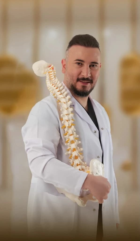
Lead specialist
Neurosurgeon, neurospine
Brain & Spine Surgery
17+ years experience
DHA License No: 20305968-001
View full profileDiagnosis → severity classification → treatment planning. No spinal stenosis treatment in Dubai is initiated without confirmed imaging and neurological assessment at DRFK.
| Diagnostic Tool | Spinal Stenosis Application |
|---|---|
| MRI Lumbar / Cervical Spine | Gold standard, canal diameter, ligamentum flavum, root compression, cord signal |
| CT Spine | Bony detail, osteophytes, foraminal narrowing, facet hypertrophy |
| Standing Flexion-Extension X-Ray | Dynamic instability, fusion need alongside decompression |
| NCS / EMG | Root level and severity, surgical level in multi-level disease |
| Walking Tolerance Assessment | Maximum distance before symptoms, severity grading and response monitoring |
Treatment decision flow: Mild → physiotherapy + laser | Moderate → injections + physio | Severe / progressive → surgical decompression.
Physiotherapy (Dr. Amina Zafar): core stabilisation, lumbar flexion-biased exercises, postural correction, aquatic therapy where available. Candidates: mild stenosis, moderate stenosis as bridge, all post-surgical rehab. Expected: improved walking tolerance within 6–12 weeks.
Laser therapy: reduces facet and ligamentous inflammation alongside physiotherapy.
Epidural steroid injection (Op. Dr. Fatih Kırar, fluoroscopy-guided): best for neurogenic claudication and cervical arm radiculopathy; walking improvement in 1–2 weeks, effect 4–12 weeks. Choose over surgery when no progressive deficit, no cauda equina, and conservative care not yet exhausted.
Spinal decompression therapy: mechanical distraction adjunct for early stenosis with disc component.
Pain management medication: anti-inflammatories, neuropathic agents (pregabalin, gabapentin), muscle relaxants, structured programme, not standalone.
Op. Dr. Fatih Kırar recommends decompression when: walking severely limited; fixed neurological deficit; cauda equina; progressive deterioration despite conservative care; or 3+ months including injections without adequate functional gain.
| Treatment | Best For | Nerve Decompression | Recovery |
|---|---|---|---|
| Physiotherapy | Mild, moderate stenosis | No | 6–12 weeks |
| Laser Therapy | Inflammatory component | No | Immediate / ongoing |
| Epidural Injection | Neurogenic claudication | Indirect | 1–2 days |
| Nerve Root Block | Foraminal stenosis, level-specific | Indirect | 1–2 days |
| Laminotomy | Single/two-level, stable spine | Yes, direct | 2–4 weeks |
| Minimally Invasive Laminectomy | Multi-level, selected cases | Yes, direct | 3–6 weeks |
| Open Laminectomy | Multi-level extensive stenosis | Yes, direct | 4–8 weeks |
| Decompression + Fusion | Stenosis + instability | Yes, direct | 3–6 months |
| Treatment | Walking Improvement | Desk Return | Full Activity |
|---|---|---|---|
| Physiotherapy | 4–8 weeks | Week 1 modified | 8–12 weeks |
| Epidural Injection | 1–2 weeks | 1–2 days | 2–4 weeks |
| Laminotomy | 1–3 weeks | 2–4 weeks | 6–8 weeks |
| Laminectomy | 2–4 weeks | 4–8 weeks | 8–12 weeks |
| Decompression + Fusion | 4–8 weeks | 6–10 weeks | 3–6 months |
Lead specialist
Neurosurgeon, neurospine
Brain & Spine Surgery
17+ years experience
DHA License No: 20305968-001
Op. Dr. Fatih Kırar, Full profileDHA-licensed spine and neurosurgery specialist, 17+ years. Complex lumbar and cervical stenosis from epidural injection and endoscopic decompression through multi-level laminectomy and fusion.

Lead specialist
Specialist Radiology
Diagnostic Imaging & Interventional Radiology
25+ years experience
DHA License No: 00219919-003
Whole Spine MRI, Dr. Vivek KapoorDr. Vivek Kapoor leads spinal stenosis diagnostic imaging at DRFK: whole spine and regional MRI, CT, and NCS with rapid reporting for severity classification and surgical planning.
25+ Years | DHA-licensed Specialist Radiology | Diagnostic Imaging & Interventional Radiology
Cost depends on modality and levels treated. Physiotherapy, injections, and indicated decompression are covered by most UAE plans including GlobeMed, pre-authorisation managed by our team.
Call +971 4 882 1015 anytime. WhatsApp DRFK Neurosurgery: +971 58 841 43 34.
Canal diameter, levels, instability, and cord signal.
Dr. Amina Zafar, flexion-biased physio, pacing, core work.
Inflammation control and mechanical adjuncts.
Op. Dr. Fatih Kırar, fluoroscopy-guided.
Laminotomy, endoscopic or open laminectomy when indicated.
When instability coexists with stenosis.
Cycling or leaning on a cart often feels easier than long upright walking, report patterns to your doctor
Avoid high-force manipulation if you have numbness or weakness
Weight control and smoking cessation support spinal circulation
Bladder or bowel change, call 999 immediately
Medically reviewed by Op. Dr. Fatih Kırar | DRFK Turkish Medical Center, Jumeirah 3, Dubai | Last updated: April 2026
Every day: 9:00 AM – 6:00 PM
Villa 2 Al Athar Street, Jumeirah 3, Dubai, United Arab Emirates