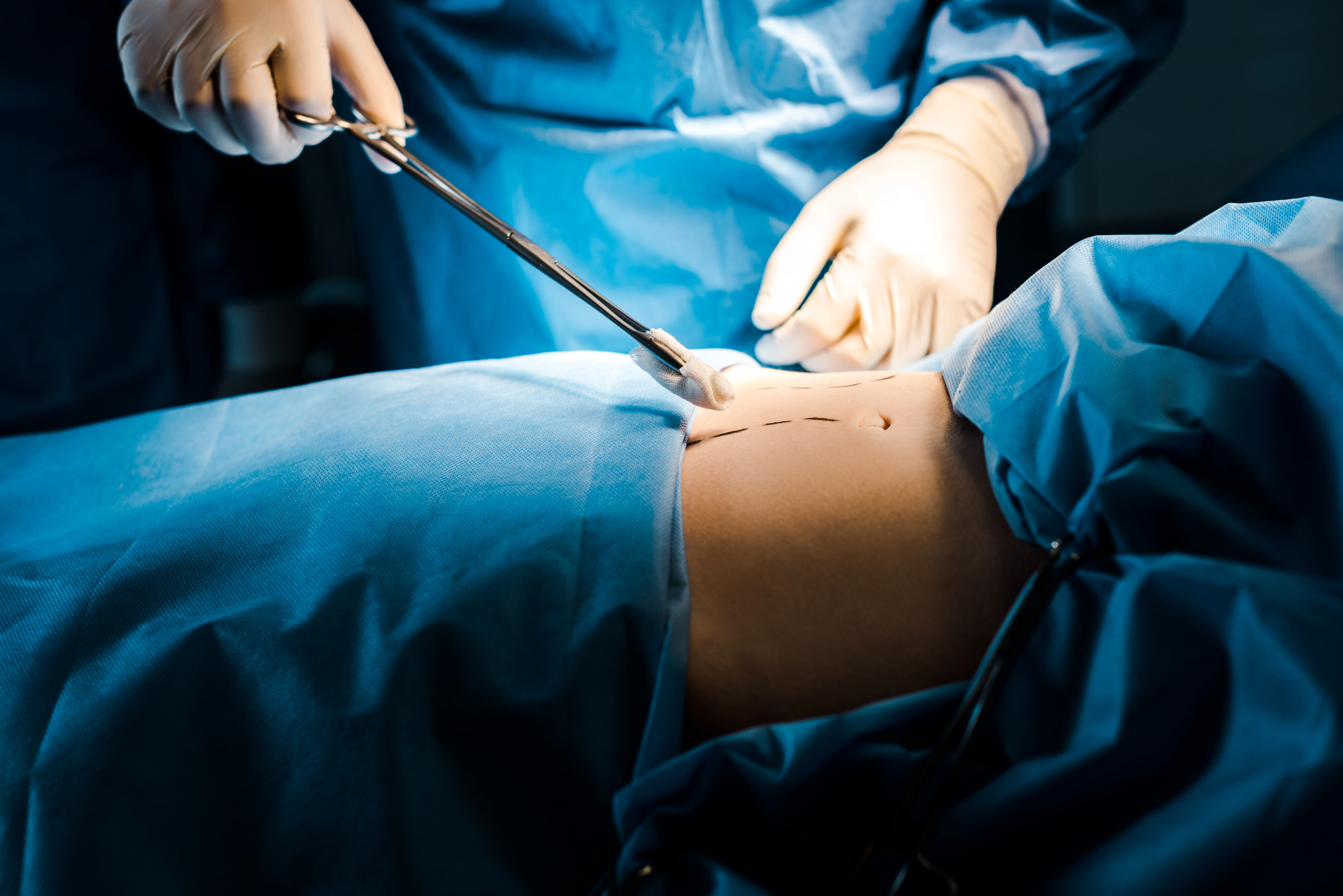
Laminotomy surgery in Dubai at DRFK Turkish Medical Center is performed by Op. Dr. Fatih Kırar, DHA-licensed spine and neurosurgery specialist (17+ years). Laminotomy removes a portion of the vertebral lamina to widen the spinal canal, decompress nerve roots, and relieve pain, numbness, and weakness from stenosis, herniated disc, and bone spur compression, preserving more stability than full laminectomy.
DRFK Turkish Medical Center is located at Villa 2, Al Athar Street, Jumeirah 3, Dubai. The clinic is easily accessible from Jumeirah, Umm Suqeim, Al Safa, Business Bay, and Downtown Dubai within around 15 minutes via Sheikh Zayed Road, with on-site parking available.
Laminotomy surgery in Dubai at DRFK Turkish Medical Center is performed by Op. Dr. Fatih Kırar, DHA-licensed spine and neurosurgery specialist (17+ years). Laminotomy removes a portion of the vertebral lamina to widen the spinal canal, decompress nerve roots, and relieve pain, numbness, and weakness from stenosis, herniated disc, and bone spur compression, preserving more stability than full laminectomy.
Unlike laminectomy, which removes the entire lamina, laminotomy removes only the bone required to decompress the nerve, preserving spinal stability and reducing post-surgical instability risk for focal single- or two-level stenosis.
The lamina is the posterior bony arch of each vertebra, the roof of the spinal canal housing the cord and nerve roots. In stenosis, the lamina, ligamentum flavum, and facets thicken and narrow the canal.
Laminotomy removes a precisely calculated portion of lamina, creating a decompressive window that relieves radiating pain, numbness, and weakness from nerve compression.
Critical distinction: laminotomy preserves structural integrity by leaving the remainder of the lamina, spinous process, and facet joint intact, the primary advantage over full laminectomy for single- or two-level stenosis without instability.
Spinal stenosis narrows the canal and compresses nerve roots → sciatica, neurogenic claudication, leg weakness on walking relieved by sitting and forward flexion.
Laminotomy removes a portion of lamina → decompresses the canal → relieves nerve pressure and ischaemic/mechanical injury within the narrowed canal.
Spinal decompression surgery encompasses all procedures that create space for compressed neural structures. Laminotomy is one type within this family.
| Procedure | Bone removed | Stability | Recovery | Best for |
|---|---|---|---|---|
| Laminotomy | Partial lamina | Preserved | Faster | Single/two-level stenosis |
| Laminectomy | Full lamina | Reduced | Moderate | Multi-level stenosis |
| Foraminotomy | Foraminal bone | Preserved | Faster | Nerve exit narrowing |
| Discectomy | Herniated disc | Preserved | Fastest | Disc herniation |
| Decompression + fusion | Lamina + instrumentation | Restored surgically | Longer | Stenosis + instability |
Step 1, Symptoms: radiating leg pain, claudication, numbness, weakness.
Step 2, Diagnosis: MRI confirms compression; NCS confirms level.
Step 3, Conservative: physiotherapy, laser, epidural injection 6–12 weeks minimum.
Step 4, Surgery: if conservative care fails and walking/function is significantly limited.
Delayed surgery: prolonged compression lowers probability of complete neurological recovery, urgency assessed at every pre-surgical consultation.
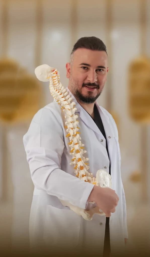
Lead specialist
Neurosurgeon, neurospine
Brain & Spine Surgery
17+ years experience
DHA License No: 20305968-001
View full profile| Diagnostic tool | Laminotomy application |
|---|---|
| MRI lumbar/cervical spine | Stenosis level, root compression, cord signal |
| CT spine | Osteophytes, facet overgrowth, foraminal narrowing |
| Standing X-ray flexion-extension | Dynamic instability, confirms laminotomy without fusion is safe |
| NCS / EMG | Root level and severity for single-level planning |
Prone positioning; general anaesthesia, Dr. Zahra Ghafari; IONM electrodes placed.
2–3 cm posterior incision; tubular retractor dilates muscle without division.
Operating microscope, high-magnification view of lamina and ligamentum flavum.
High-speed drill and Kerrison rongeur create decompressive window; facet preserved.
Root visualised free, pulsating normally; residual bone or disc removed.
Absorbable sutures; mobilise 4–12 hours; discharge day 1–2 most cases.
Clinical principle at DRFK: anatomy and symptoms determine technique, laminotomy when focal decompression achieves the objective; laminectomy when multi-level canal expansion is required.
| Feature | Laminotomy | Laminectomy |
|---|---|---|
| Bone removed | Partial window | Full lamina |
| Posterior stability | Preserved | Reduced |
| Instability risk | Low | Higher, may need fusion |
| Incision | Smaller | Larger |
| Recovery | Faster | Slightly longer |
| Hospital stay | 1–2 days | 2–4 days |
| Best for | Focal single-level | Multi-level extensive |
| Fusion required | Rarely | Sometimes |
Non-surgical decompression reduces intradiscal pressure for early disc irritation. Laminotomy is required when compression is from bone and ligamentum flavum overgrowth that conservative care cannot reverse. MRI-confirmed stenosis may improve temporarily with non-surgical care but bony narrowing remains until surgically addressed.
Is laminotomy permanent? Bone removed does not regenerate, immediate decompressive effect is permanent. New bone formation, osteophyte growth, and ligamentum flavum hypertrophy can occur over time at adjacent or operated levels, core strengthening and monitoring are recommended.
| Phase | Timeline | What to expect |
|---|---|---|
| Day of surgery | 0 | 1–2 hrs; mobilise 4–12 hrs |
| Day 1–2 | Post-op | Discharge; walking encouraged |
| Week 1–2 | Early | Desk possible week 2; no heavy lifting |
| Week 2–4 | Physio | Dr. Amina Zafar, core strengthening |
| Week 4–6 | Active | Full desk; light exercise |
| Week 6–12 | Full | Unrestricted activity; return to sport |
| 3 months | Review | MRI/X-ray if symptoms persist |
Lead specialist
Neurosurgeon, neurospine
Brain & Spine Surgery
17+ years experience
DHA License No: 20305968-001
Op. Dr. Fatih Kırar, Full profileDHA-licensed spine and neurosurgery, 17+ years. Principle: least bony removal necessary for complete neurological decompression.
Cost depends on levels decompressed and complexity. Laminotomy for documented neurological deficit is covered by most UAE plans, pre-authorisation managed by our team.
Confirm compression level and instability screening.
2–3 cm incision, tubular retractor, microscope-guided.
Targeted decompressive window; facet preserved.
Continuous monitoring, Dr. Zahra Ghafari anaesthesia.
Dr. Amina Zafar, core strengthening from week 2.
Complete 6–12 weeks structured conservative care before surgical decision unless urgent neurology
Report new leg weakness or bladder change immediately
Walking from day 1 accelerates recovery
Finish full physiotherapy programme to reduce recurrence risk
Bring MRI and NCS reports to consultation
Medically reviewed by Op. Dr. Fatih Kırar | DRFK Turkish Medical Center, Jumeirah 3, Dubai | Last updated: April 2026
Every day: 9:00 AM – 6:00 PM
Villa 2 Al Athar Street, Jumeirah 3, Dubai, United Arab Emirates