A brain MRI is a non-invasive diagnostic imaging procedure that uses magnetic fields and radio waves to create detailed images of the brain and surrounding structures. It may help evaluate neurological symptoms, detect abnormalities such as tumours or stroke, assess brain injuries, and support diagnosis and treatment planning without using ionizing radiation. At DRFK Turkish Medical Center, Jumeirah 3, scans are personally reported by Dr. Vivek Kapoor, DHA-licensed Specialist Radiologist.
DRFK Turkish Medical Center is located at Villa 2, Al Athar Street, Jumeirah 3, Dubai. The clinic is easily accessible from Jumeirah, Umm Suqeim, Al Safa, Business Bay, and Downtown Dubai within around 15 minutes via Sheikh Zayed Road, with on-site parking available.

Lead specialist
Specialist Radiology
Diagnostic Imaging & Interventional Radiology
25+ years experience
DHA License No: 00219919-003
Dr. Vivek KapoorA brain MRI is a diagnostic imaging procedure that uses magnetic fields and radio waves to create detailed images of the brain and surrounding structures. It is commonly used to evaluate neurological symptoms, investigate abnormalities, and assist healthcare professionals in diagnosis and treatment planning. At DRFK Turkish Medical Center, every brain MRI is reviewed by Dr. Vivek Kapoor, a Specialist Radiologist with over two decades of experience in diagnostic imaging.
The scan works without radiation. A strong magnetic field and radio waves cause signals from the body's tissues, which a computer reconstructs into highly detailed images. Because magnetic resonance imaging shows soft tissue so clearly, it is particularly valuable for the brain, where fine structural detail matters.
The brain is a complex structure, and a brain MRI visualises its parts in detail to support accurate diagnosis. Key structures include the cerebrum, cerebellum, brainstem, ventricles, pituitary gland, optic nerves, and the surrounding meninges and blood vessels, alongside the white matter and gray matter that carry and process signals.
Understanding this anatomy explains why an MRI scan of the brain is so useful. Different conditions affect different structures, so detailed visualisation of each region allows a neuroradiologist to localise a problem and support the clinical picture, which is then correlated with symptoms by the treating doctor.
A brain MRI is recommended when neurological symptoms or clinical findings suggest a condition that needs detailed imaging of the brain. It follows clinical assessment rather than replacing it, helping to investigate the cause of specific symptoms.
Common reasons a brain scan may be advised include:
Identifying the right indication is important, which is why imaging is requested in the context of a neurological consultation and the symptom pattern.
A brain MRI provides detailed visualisation of brain structures, blood vessels, white matter, gray matter, and surrounding tissues. It may assist in evaluating conditions such as brain tumours, stroke, multiple sclerosis, inflammatory disorders, traumatic injuries, and structural abnormalities, depending on the clinical context and imaging protocol used.
Among the conditions an MRI scan brain study can help evaluate are tumours, areas of stroke or bleeding, demyelinating disease such as multiple sclerosis, infections, aneurysms and vascular abnormalities, hydrocephalus, and neurodegenerative changes. The findings are always interpreted alongside the patient's symptoms, since imaging supports diagnosis rather than standing alone.
A brain MRI is one of the most important tools for evaluating a suspected brain tumour, showing its location, size, and relationship to surrounding structures. A brain tumour MRI typically uses contrast to highlight abnormal tissue and its blood supply, helping to characterise the lesion.
A common question is whether MRI without contrast can detect a brain tumour. Non-contrast MRI can often identify a mass, but contrast frequently adds important information about its nature and margins. It is also worth noting that not all lesions behave identically on imaging, so findings are interpreted by a radiologist and correlated clinically, with brain tumour evaluation coordinated with the neurosurgical team where needed.
Whether contrast is used depends on the clinical question the scan is meant to answer. A non-contrast brain MRI is sufficient for many indications, while a contrast MRI using gadolinium is added when more information is needed about tumours, infection, inflammation, or vascular structures.
Contrast helps highlight areas where the blood-brain barrier is disrupted, making certain abnormalities clearer. The decision to use contrast is made based on the suspected condition and safety screening, and it is explained to the patient beforehand. This judgement is part of the protocol planning Dr. Vivek Kapoor applies to each case.
A brain MRI can help assess brain damage from causes such as traumatic brain injury, stroke, white matter changes, and oxygen deprivation, depending on the type and timing of injury. It visualises structural change and signal abnormalities that may reflect tissue damage.
A brain MRI can also contribute to evaluating certain vision problems, because it images the optic nerves, pituitary gland, and related pathways. Conditions affecting these structures, such as optic nerve disorders or pituitary lesions, may be assessed on imaging, then correlated with an ophthalmology or neurology assessment for a complete picture.
A brain MRI is performed with the patient lying still inside the MRI scanner while images are acquired. Before the scan, a safety screening checks for implanted devices, metal, or conditions that need extra evaluation, since the magnetic field interacts with certain materials.
Preparation is usually straightforward. Patients are asked about implants and previous surgery, and for most brain MRI studies normal eating and medication can continue unless told otherwise; if sedation or specific contrast preparation is needed, the imaging team gives instructions in advance. Remaining still during the scan is important for clear images.
A brain MRI is generally considered a safe, non-invasive imaging technique because it does not use ionizing radiation. Side effects are uncommon, though the scanner is noisy and some people find the enclosed space uncomfortable, and where gadolinium contrast is used it is generally well tolerated with rare reactions.
Safety depends on screening. Certain implanted medical devices, metallic objects, or specific health conditions may require additional evaluation before imaging, which is why a safety check is performed before every examination. Patients are encouraged to share their full medical history so the scan is planned safely.
Brain MRI results are interpreted by a radiologist who examines the images and produces a report, which is then correlated with the patient's symptoms by the treating doctor. At DRFK, Dr. Vivek Kapoor personally reviews and reports studies rather than outsourcing interpretation, with same-day reporting pathways in standard cases.
Timing depends on the protocol and complexity, with many reports available quickly and integrated follow-up arranged when further evaluation is recommended. Because imaging supports rather than replaces clinical judgement, specialist interpretation and correlation with symptoms are essential to an accurate diagnosis.
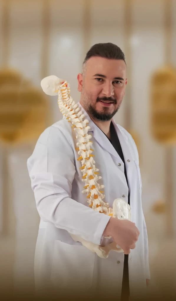
Lead specialist
Neurosurgeon, neurospine
Brain & Spine Surgery
17+ years experience
DHA License No: 20305968-001
Op. Dr. Fatih KırarDRFK Turkish Medical Center is a DHA-licensed facility in Jumeirah 3 offering in-house diagnostic imaging with personal specialist reporting rather than outsourced interpretation. Brain MRI studies are reported by Dr. Vivek Kapoor with same-day pathways in standard cases, and findings can be reviewed directly with the neurosurgical team led by Op. Dr. Fatih Kırar when clinically relevant, giving an integrated pathway from scan to specialist.
The service is available to patients across Jumeirah, Umm Suqeim, Al Safa, Al Wasl, City Walk, Business Bay, Downtown Dubai, DIFC, Dubai Marina, Palm Jumeirah, JBR, Emirates Hills, Dubai Hills, and Arabian Ranches, with multilingual support for international patients and self-referred booking available.
Medically reviewed by Dr. Vivek Kapoor, Specialist Radiology, DRFK Turkish Medical Center, Jumeirah 3, Dubai.
Every day: 9:00 AM – 6:00 PM
Villa 2 Al Athar Street, Jumeirah 3, Dubai, United Arab Emirates