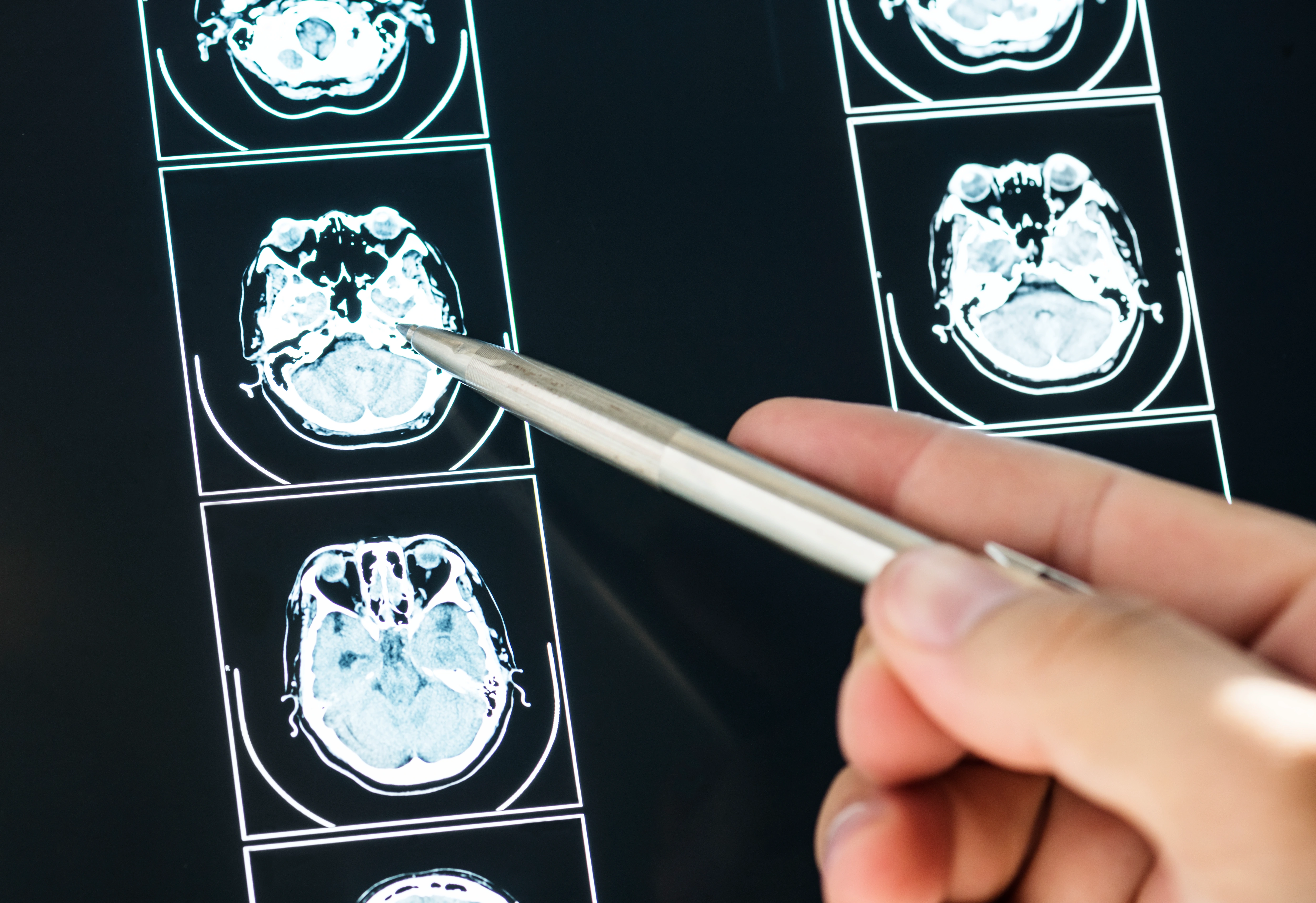
Primary and secondary brain tumours need a team combining precise imaging, neurosurgical expertise, and supportive care. At DRFK Turkish Medical Centre — among Dubai's neurosurgery centres — we help you understand your diagnosis clearly, discuss surgery versus focused radiation, monitor therapy side effects, and plan recovery under Op. Dr. Fatih Kirar, with 17+ years of experience and more than. Complex cases are reviewed by a multidisciplinary team before any final recommendation. The centre is at Villa 2, Al Athar Street, Jumeirah 3, Dubai — around 15 minutes from Jumeirah, Umm Suqeim, Al Safa, Business Bay, and Downtown via Sheikh Zayed Road, with on-site parking.
DRFK Turkish Medical Center is located at Villa 2, Al Athar Street, Jumeirah 3, Dubai. The clinic is easily accessible from Jumeirah, Umm Suqeim, Al Safa, Business Bay, and Downtown Dubai within around 15 minutes via Sheikh Zayed Road, with on-site parking available.
New or worsening headaches, seizures, personality or behavioural change, visual disturbance, progressive limb weakness, or speech difficulty need urgent assessment. We prioritise time-sensitive work-up when there is raised suspicion of mass effect.
Urgent assessment
New gradually worsening headache, seizures, personality change, visual changes, limb weakness, or speech difficulty — these cases receive immediate appointment priority when a compressive mass is suspected. Emergency: sudden consciousness deterioration, sudden severe headache, or a seizure lasting more than 5 minutes — call 999 immediately.
The pathway starts with a detailed history, full neurological examination, and careful MRI review. Depending on tumour type, location, and grade, neurosurgery, oncology, and rehabilitation teams are involved in the decision. The goal is clear: a plan that achieves maximum treatment effectiveness while preserving neurological function and quality of life — a good outcome is not just tumour removal, but removal with the patient able to live, work, and communicate.
Beyond acute treatment, we support medication titration, structured corticosteroid weaning, seizure control, rehabilitation referrals, psychological and family counselling, and scheduled imaging to detect recurrence or progression early.
| Type | Classification | Common Examples | Typical Approach |
|---|---|---|---|
| Primary benign | Originates in the brain — non-cancerous | Meningioma, schwannoma | Complete resection often; observation for small silent lesions |
| Primary malignant | Originates in the brain — cancerous | Gliomas (graded) | Maximal safe resection followed by radiation and chemotherapy |
| Metastatic (secondary) | Spreads from cancer elsewhere | Lung, breast, kidney, or skin metastases | Surgery or focused radiation with systemic therapy as indicated |
Structured imaging review focuses on two critical questions: Is the tumour resectable? And how close is it to sensitive functional areas?
| Diagnostic Tool | Role |
|---|---|
| Contrast-enhanced MRI | Gold standard: characterises tumour, margins, and relation to eloquent areas |
| Functional MRI (fMRI) | Maps motor and language centres before surgery |
| MR spectroscopy (MRS) | Metabolic fingerprint supporting grade estimation before biopsy |
| CT scan | Rapid assessment for haemorrhage and emergencies |
| Biopsy | Confirms tumour type and grade histologically — basis for adjuvant planning |
Biopsy to confirm diagnosis, decompression to relieve pressure, or resection to the greatest safe extent — using neuronavigation, continuous intraoperative monitoring (IONM), and 5-ALA fluorescence for high-grade tumours when indicated.
Full coordination of radiation therapy according to tumour type and grade — alternative or complement to surgery in selected cases.
Coordination of chemotherapy and targeted therapies with careful monitoring of side effects.
Not every tumour needs immediate intervention — some slow-growing, silent meningiomas are managed with interval imaging. We explain clearly when resection becomes the right decision.
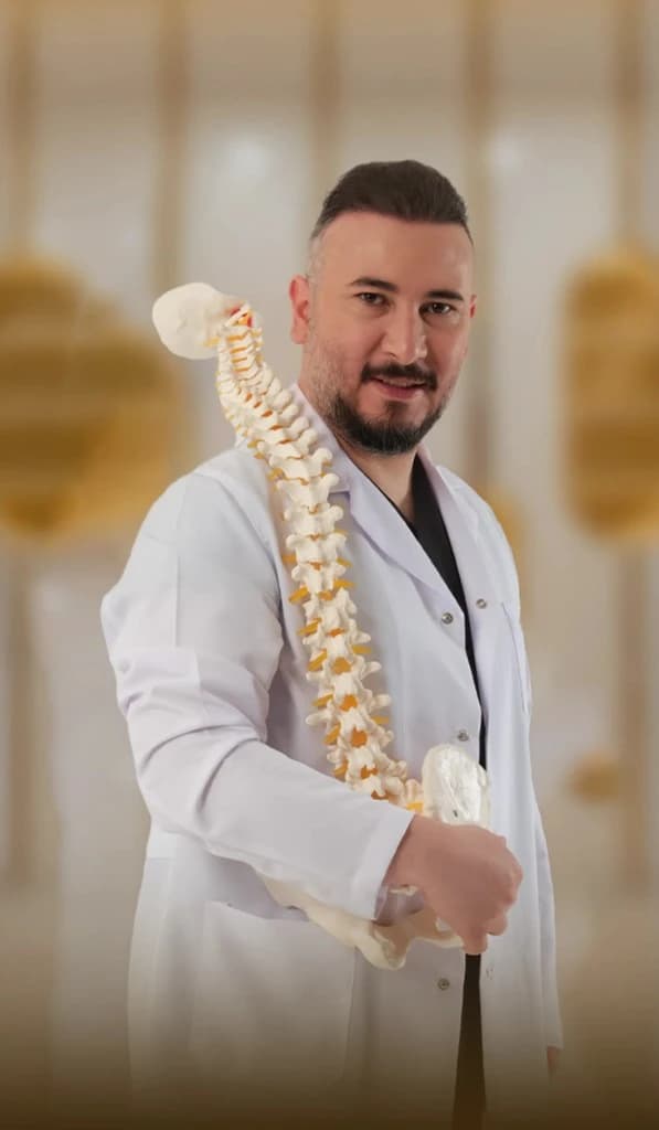
Lead specialist
Neurosurgeon, neurospine
Brain & Spine Surgery
17+ years experience
DHA License No: 20305968-001
Full profile — Op. Dr. Fatih KirarOp. Dr. Fatih Kirar leads the brain tumour pathway at DRFK — from imaging review to surgery to coordination of adjuvant therapy — with an approach that always balances treatment effectiveness and neurological safety.
Cost depends on tumour type, treatment plan, and duration of care; a predetermination estimate can be requested after assessment. Insurance coverage varies by plan — the DRFK team supports prior-authorisation processes. Please confirm your plan details with patient services.
Emphasis on resectability and eloquent-area risk for every case.
Biopsy, decompression, or resection with neuronavigation and IONM when indicated.
brain tumour surgery DubaiRadiation and/or chemotherapy aligned with the oncology team; symptom monitoring.
Structured dosing and taper to minimise oedema and protect against breakthrough seizures.
Physiotherapy, occupational therapy, and speech therapy as needed for recovery.
Interval imaging schedule tailored to your tumour profile.
Bring a family member to consultation — decisions are complex and a second listener helps
Keep a complete folder of reports and disc/USB copies of imaging; comparing scans over time reveals progression
Report any sudden neurological change immediately — new weakness, seizure, or consciousness change
Page content reviewed and approved by Op. Dr. Fatih Kirar, DHA-Licensed Specialist Surgeon in Neurosurgery | DRFK Turkish Medical Centre, Jumeirah 3, Dubai | Last updated: July 2026. Information is for education and does not replace personalised medical advice.
Every day: 9:00 AM – 6:00 PM
Villa 2 Al Athar Street, Jumeirah 3, Dubai, United Arab Emirates