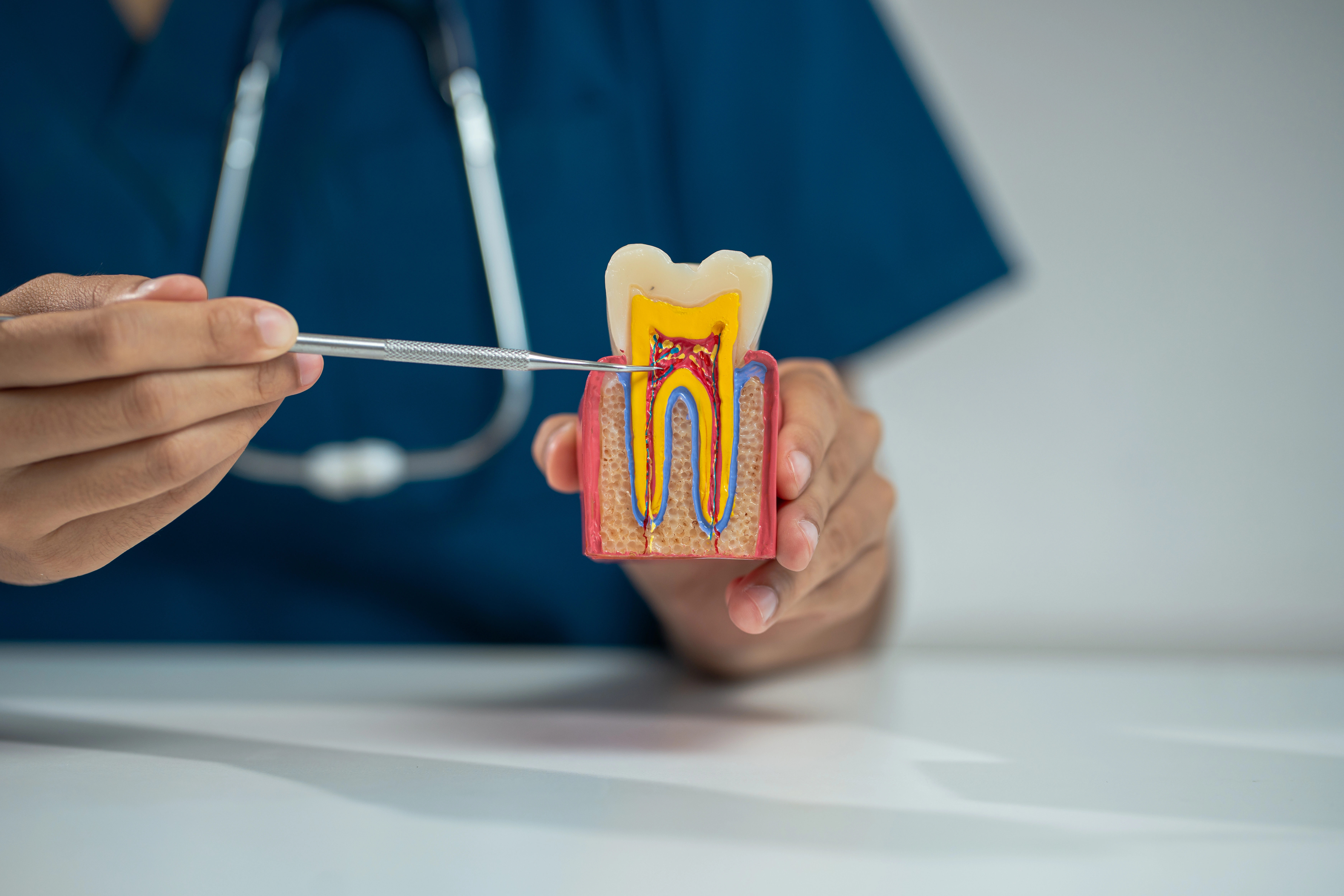
Root canal treatment in Dubai at DRFK Turkish Medical Center couples microscope-guided precision with compassionate anaesthesia, typically completed by Dr. Raneen Essale in one visit when clinically appropriate. Toothache, swelling, or imaging-confirmed pulp disease no longer mean sacrificing teeth unnecessarily.
DRFK Turkish Medical Center is located at Villa 2, Al Athar Street, Jumeirah 3, Dubai. The clinic is easily accessible from Jumeirah, Umm Suqeim, Al Safa, Business Bay, and Downtown Dubai within around 15 minutes via Sheikh Zayed Road, with on-site parking available.
Microscopic magnification, rotary instrumentation, rubber dam isolation, and seamless restorative partnerships differentiate specialist-led care.
Root canal therapy removes infected or necrotic pulp tissue from every canal, disinfects the system, and seals it permanently, preserving tooth structure while eliminating infection.
Treatment at DRFK is microscope-guided with rotary instrumentation under local anaesthesia; many cases finish in one appointment.
Step 1, Diagnosis and CBCT assessment to map canal anatomy and periapical pathology when needed.
Step 2, Local anaesthesia with comfort-focused delivery; supplementary techniques for difficult pulpal cases.
Step 3, Rubber dam isolation to ensure a sterile field and prevent contamination.
Step 4, Microscope-guided access and canal identification including accessory canals.
Step 5, Rotary NiTi instrumentation with sodium hypochlorite and EDTA irrigation protocols.
Step 6, Warm gutta-percha obturation to seal canal systems against reinfection.
Step 7, Post-endodontic restoration planning with timely crown protection.
Clinical assessment plus imaging, CBCT when anatomy demands mapping canals and lesion extent.
Topical anaesthetic precedes slow injection; rubber dam isolation maintains sterility.
Operating microscope reveals accessory canals; rotary NiTi shaping plus antimicrobial irrigation.
Warm gutta-percha compaction seals canals, CAD-CAM crown planned promptly to reduce fracture risk.
Mild tenderness for 2-5 days is common and usually controlled with prescribed anti-inflammatory medication.
Avoid heavy chewing on the treated side until the definitive crown is fitted. Delaying crown placement raises fracture risk.
Saving natural teeth preserves bone volume, neighbouring contacts, and efficiency versus extraction followed by implant therapy when salvage remains realistic.
| Root canal + crown | Extraction + implant | |
|---|---|---|
| Preserves natural tooth | Yes | No |
| Jawbone preservation | Yes | Requires implant for stimulation |
| Function | Close to natural tooth function | Restored after extended treatment timeline |
| Cost comparison | Lower | Higher |
| Preferred option | First choice when tooth is salvageable | When tooth is not salvageable |
Periapical / CBCT mapping prior to instrumentation.
Chemomechanical disinfection with rotary NiTi systems.
Removal of prior fills with microscopic retreatment pathways.
CAD-CAM crowns planned promptly after canal sealing.
Complete antibiotic courses only when prescribed.
Alert clinicians if swelling worsens or fever develops.
Wear temporary crowns carefully, sticky foods dislodge temporaries.
Discuss night guards if parafunction threatens ceramics.
Follow recall intervals for occlusion checks.
Medically reviewed by Dr. Raneen Essale | DRFK Turkish Medical Center, Dubai | Last updated: April 2026
Every day: 9:00 AM – 6:00 PM
Villa 2 Al Athar Street, Jumeirah 3, Dubai, United Arab Emirates